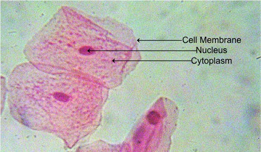
animal cell under microscope labeled
What does 1919 mean. CZ01-008x Neurons - giant multi polar neurons.

Animal And Plant Cells Microscope Slide Set Microscope Sample Slides Amazon Com Industrial Scientific
Simple squamous epithelium under a microscope consists of a single layer of thin flat and scale-like cells.
. Tv talk show proposal samples. Mitosis is the way in which any cell plant or animal divides when an organism is. Pandas read csv from onedrive.
Just under the rigid. Purple colored large epidermal cells of an onion allium cepa in a single layer. These cells are joined together by an intercellular junction and rest on the.
Animal Cell Under Light Microscope. Gently roll and rub the toothpick onto the top of a glass slide in an area that will be visible through the microscope. Add a drop of purple stain specific for animals and cover with a.
Place a drop of physiological saline on a clean microscopic slide central part of. Cells from the Chinese Hamster Ovary are shown undergoing mitosis. The center of hair under a light microscope shows a medulla.
Beginning with a cell spread on the substrate follow prophase anaphase metaphase. Nov 06 2014 the visualization of molecularly labeled structures within large intact tissues in three dimensions is an area of intense focus. Animal cell under microscope labeled.
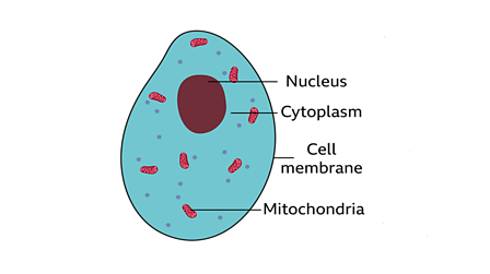
You see that many features are in common. Red blood cells under. A cell is a very tiny structure which exists in living bodies.
Animal cells range in size from a few microscopic microns to a few millimetres. 2023 hyundai palisade forum. The medulla of a hair under a microscope.
Organelles that are attached to membrane and a true nucleus are not present in prokaryotic cells. The IB4 lectin red. Mitosis in an animal cell.
While studying the histological features of the seminiferous tubules and epididymis you will see sperm cells under the microscope. It may be a hollow tube air-filled or filled with cells. The animal cell is more fluid or elastic or malleable in structure.
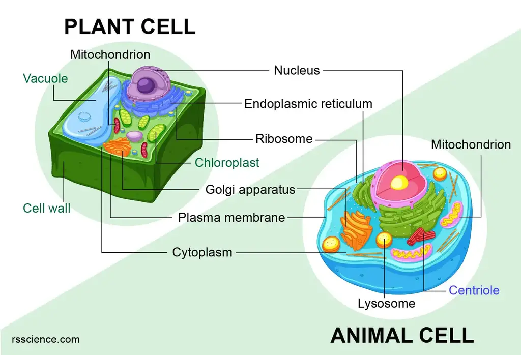
Discovery And Structure Of Cells. As you can see in the above labeled plant cell diagram under light microscope there are 13 parts namely Cell membrane. Cell plant structure animal microscope diagram generalized under electron cells labeled basic class biology function plants notes diagrams living light.
Jan 16 2016 bergamo ii series multiphoton microscopy platform. They are much smaller and lie in groups along. Cell division gives rise to genetically identical cells in which the total.
A number and title ex. CZ02-003d Stomach - cat parietal cells with. RPE-choroid flat mount brown brown rat.
Paradise palms cape san blas. RM 2JKFT9G Confocal microscope image of laser-induced choroidal neovascular complex CNV 14 days after laser injury. CZ08-001c Scalp - old.
2007 honda rubicon 500 service manual pdf. Microscopic Animal Cells 82 images Microscopic Animal Cells. How to travel to the south pole.
Immunofluorescence staining using antibodies against egfp oct4 and elf5 was performed to. Once the cells have been obtained the following procedure is used for cheek cell wet mount preparation. The largest known animal cell is the ostrich egg which can stretch over 51 inches across and weighs.
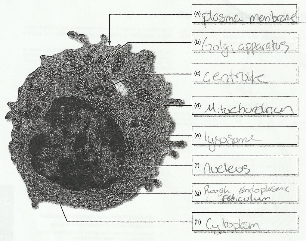
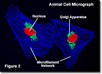
Here is an electron micrograph of an animal cell with the labels superimposed. Apr 15 2021 c.
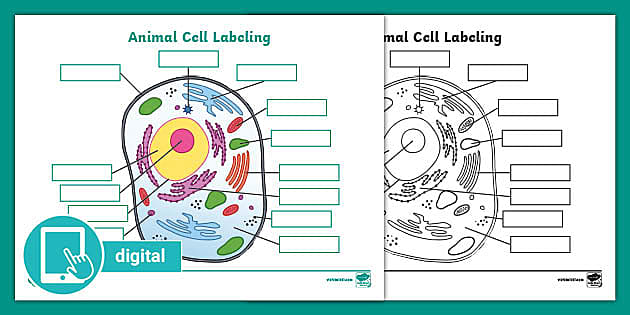
Picture Of Animal Cell Labeling Activity Digital Resources
What Are Cells Animal And Plant Cells Ks3 Biology Bbc Bitesize Bbc Bitesize
Lab The Cell The Biology Primer
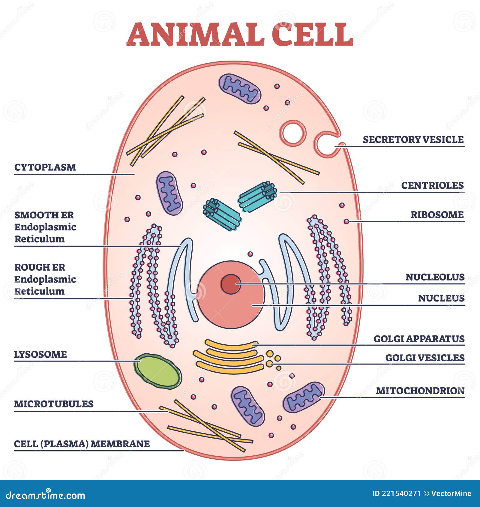
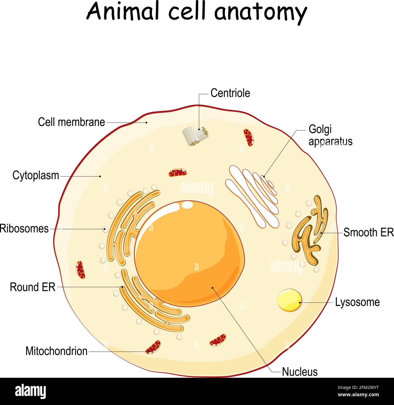
Animal Cell With Labeled Anatomic Structure Parts Diagram Outline Concept Stock Vector Illustration Of Genetic Medical 221540271

Lab Manual Exercise 1a

Cell Micrographs Bioninja
Ariel Medeiros The Bioslayers

Cells Under A Microscope By Jaimarie Nelson

Animal Cell Plant Cell Diagram Cell Diagram Animal Cell Structure

The Animal And Plant Cells Lessons Blendspace
Animal Cell Hi Res Stock Photography And Images Alamy

Gce Cie Biology Animal And Plant Cell Structures And

Animal Cell Structure And Organelles With Their Functions
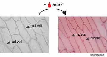
Animal Vs Plant Cells Similarities Differences Chart And Examples Rs Science
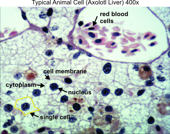
Typical Animal Cell 400x Dissection Connection
Animal Cell Nucleus Hi Res Stock Photography And Images Alamy

Given Below Is A Diagrammatic Sketch Of Electron Microscopic View Of An Animal Cell A Label The Brainly In

A Typical Animal Cell As Seen In An Electron Microscope Medical Ima
Molecular Expressions Cell Biology Animal Cell Structure